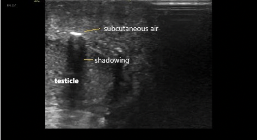
A 60 year-old male with history of poorly controlled diabetes, congestive heart failure, chronic kidney disease, and a prior MI presented as a transfer from an outside hospital for surgical management of suspected necrotizing fasciitis/Fournier’s gangrene. He reported symptoms of diarrhea, nausea, vomiting, and severe pain in the scrotum and perineum for several days. This began after he sustained a small cut to the area. He denied fevers, urinary discharge, respiratory symptoms, chest pain, but did endorse chills and night sweats…
Read MoreWhen a patient presents to the emergency department with atraumatic monoarticular arthritis, it is important to rapidly determine the etiology because septic arthritis is a medical emergency that can lead to significant morbidity and mortality…
Read MoreA 32-year-old female with no significant past medical history presents to the emergency department with complaints of nausea, vomiting, and bloating. The patient reports her last menstrual period was about 2 months prior to presentation. She was evaluated about one-month ago, at which time she was told that a pelvic ultrasound revealed an intrauterine gestation sac without a fetal pole or yolk sac, suspicious for early pregnancy failure. The patient subsequently developed vaginal bleeding, associated with abdominal cramping, nausea, and vomiting. She interpreted these symptoms as a miscarriage. The patient states that the vaginal bleeding has since subsided, but she endorses continued and progressive nausea, vomiting, and bloating. She is unable to tolerate oral intake. She denies fever, headache, abdominal pain, back pain, vaginal discharge/odor/pain, dysuria, or diarrhea…
Read MoreWe use ultrasound for a multitude of procedures in the emergency department, including central and peripheral venous access, abscess drainage, and joint aspiration. Less common, but just as useful, is the use of ultrasound to help identify landmarks when performing a lumbar puncture (LP). While the traditional method of LP is based on palpating anatomical landmarks, this is becoming increasingly difficult as the patient population becomes more obese. It may also be difficult in patients who have abnormal spinal anatomy or in those who have had previous spinal surgery. When palpating anatomy is difficult, ultrasound can be a useful tool to help identify appropriate landmarks and increase your LP success rate…
Read MoreA 48 year-old female with idiopathic intracranial hypertension (IIH), with recent shunt removal, presented with increasing right eye vision loss over the last week, headache, and left-sided weakness. Her outpatient neuro-ophthalmologist referred her to the emergency department from clinic for a question of intracranial abscess, optic neuritis, or worsening IIH. She needed an MRI as soon as possible to rule-out these dangerous pathologies. A quick chart review showed that she has needed multiple central lines placed in the past, due to difficult IV access. The nurse was unable to place a peripheral IV…
Read More